Professor Raymond Phaneuf
Much of how we perceive objects has to do with the way that light interacts with the materials they are made from; this interaction is the basis for the field of optics. In recent years unconventional materials, containing controlled nanometer scale structures have been developed to change the interaction of light with objects in dramatic ways.
Light is a propagating electromagnetic field which oscillates in time with a well defined frequency and in space, with a corresponding wavelength; for extremely small metal particles, those much smaller than a wavelength of light (~350-700 nm in the visible) the "sea of electrons" that make metals good conductors is driven back and forth by the oscillating field, at the frequency of the incoming light. Because there is a restoring force when the electrons are moved from their normal positions, there is also a natural oscillation frequency: oscillation of the position of the sea of electrons at this frequency is called a particle plasmon. Resonance occurs if the frequency of the light matches that of the particle plasmon.
Since we can tune the natural frequency of the plasmon by changing the particle shape, we can arrange for resonance at any frequency in the visible part of the light spectrum. Why is this important? The oscillation of the charge in the particles creates stronger electric fields around the particle than those due to the incident light alone. In turn, stronger electric fields have the effect of increasing the brightness of fluorescent molecules, and these give off more photons of a different color when they are excited by the same light beam that causes the electron movement. This effect we call "nanoparticle-enhanced fluorescence." The effect is so large that when we coat an entire slide with fluorescent dye, and shine light on it, we can see (with a light microscope) the areas where we have put metal nanoparticles as clearly brighter than areas without metal nanoparticles, and measure light intensities up to several hundred times brighter than normal [1]. The amount of enhancement is based on the shape and spacing of the nanoparticles, the substrate underneath them, and the wavelength of light used.
[Figure 1. SEM/reflectance/fluorescence images of surface with different sized nanoparticles and fluorescent dye. SEM images show size and shape of the particles, the reflectance image shows where light is not absorbed by the silver particles (some sizes of nanoparticle absorb this wavelength very well, and those areas look dark), and the fluorescence image shows where dye is made brighter by nanoparticle enhancement. Dye evenly coats the entire field of view, but is visibly enhanced near the right sized nanoparticles.]
This much has been known for some time. Recently, however, we discovered that another type of charge oscillation or plasmon, one which moves along the surface of a metal can also contribute to enhancing fluorescence [2]. Ordinarily, light doesn't couple to such "surface plasmons" because of the different relationship between frequency and propagation vector for these two different types of waves. (The propagation vector k is given by 2π divided by the wavelength λ). Our results show that for light incident on a periodic array of silver nanowires we can excite a standing wave, consisting of two surface plasmons moving along the surface in opposite directions, i.e. with equal and opposite propagation vectors. Our results further show that the maximum fluorescence occurs when our dye molecules coat nanowires whose widths are equal to an integer times the wavelength associated with a surface plasmon. A width of one wavelength allows for the maximum polarization of charge at opposite sides of the wire, and the largest coupling to the incoming light. A wire width of two wavelengths gives no net polarization, and very little fluorescence. Three wavelengths across a wire gives a smaller enhance ment than one due to the smaller polarization. Using this new insight, we can predict more accurately the nanostructure array parameters for maximum fluorescent output over a range of incident light wavelengths. This will have considerable impact on the development of highly sensitive detectors for hazardous biomolecules, allowing much smaller quantities to be detected.
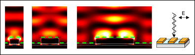
[Figure 2. Calculated electric field intensities in the area above long nanowires of silver, excited by 514 nm light. Outlines of the material interfaces, with the silver on an aluminum oxide layer over a silicon wafer, are shown as dotted lines. The nanowires are 158, 256, and 512 nm wide, respectively, and separated by distances equal to their width. The angle of observation of the material cross section, and the direction and polarization of the incident light, are demonstrated in the cartoon at the far right: the long axes of the bar is normal to the plane of the image, and only one is shown, but the calculations represent many parallel wires on the surface.]
References:
[1] Guo, S.H., S.J. Tsai, H.C. Kan, D.H. Tsai, M.R. Zachariah, and R.J. Phaneuf. The effect of an active substrate on nanoparticle-enhanced fluorescence. Adv. Mater. 2008. 20, 1424-1428.
[2] Guo, S.-H., J.J. Heetderks, H.-C. Kan, and R.J. Phaneuf. Enhanced fluorescence and near-field intensity for Ag nanowire/nanocolumn arrays: evidence for the role of surface plasmon standing waves. Opt. Express 2008. 16, 18417-18425.
Top